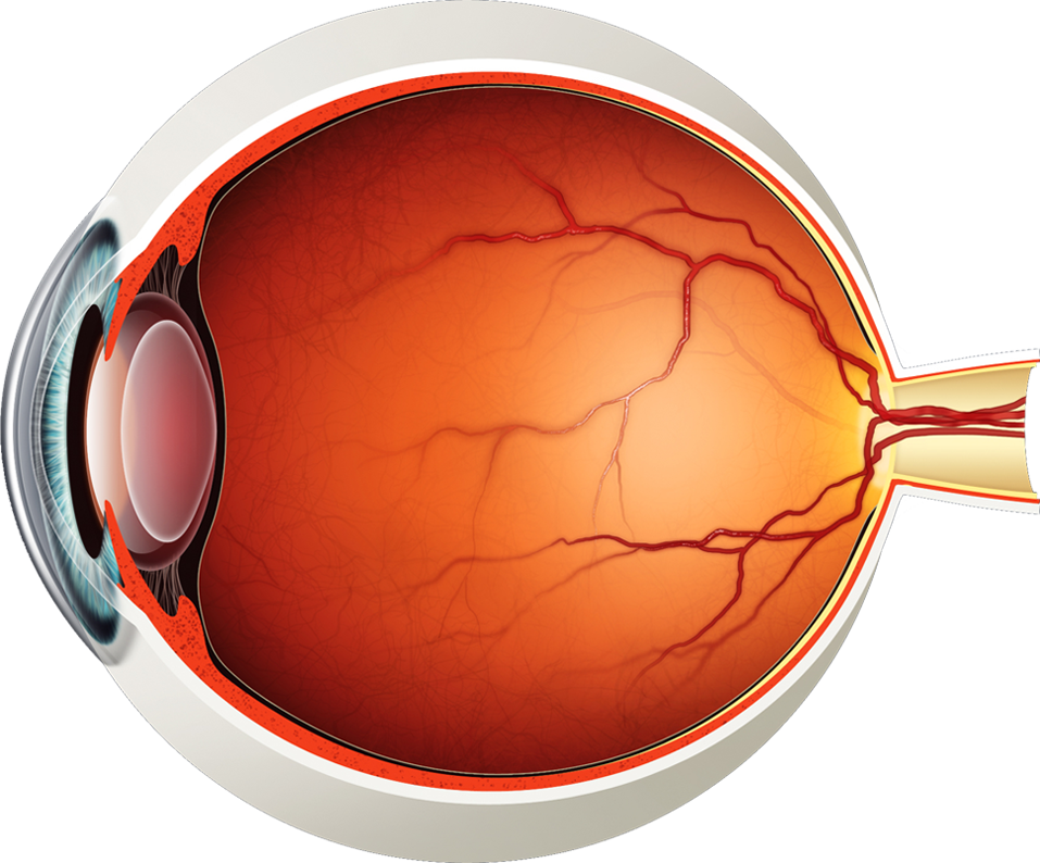
The eye – one of the most complex organisms in the human body.
It is made up of many different parts working in unison together. In order for the eye to work at its best, all parts must work well collectively.
To understand the eye and its functions, it’s important to understand how the eye works, see below diagrams for both the external eye and the internal eye.
The External Eye
Instructions
Click the parts of the eye to see a description for each. Hover the diagram to zoom.
-
Instructions
Click the parts of the eye to see a description for each. Hover the diagram to zoom.
-
Eyelid
An eyelid is a thin fold of skin that covers and protects the eye.
-
Pupil
The pupil is the opening in the centre of the iris that regulates the amount of light entering the eye. The size of the pupil determines the amount of light that enters.
-
Iris
The iris is the coloured part of the eye which surrounds the pupil. It controls light levels inside the eye, similar to the aperture on a camera. The iris contains tiny muscles that widen and narrow the pupil size. The colour, texture, and pattern of an iris are as unique as a fingerprint.
-
Sclera
The sclera is the white covering that protects the eye, commonly known as ‘the white of the eye’. Part of the sclera can be seen at the front of the eye. It is tough tissue which serves as the eye's protective outer coat.

The Internal Eye
Instructions
Click the parts of the eye to see a description for each. Hover the diagram to zoom.
-
Instructions
Click the parts of the eye to see a description for each. Hover the diagram to zoom.
-
Iris
The iris is the coloured part of the eye which surrounds the pupil. It controls light levels inside the eye, similar to the aperture on a camera. The iris contains tiny muscles that widen and narrow the pupil size. The colour, texture, and pattern of an iris are as unique as a fingerprint.
-
Lens
The lens of the eye is located behind the pupil, its purpose is to focus light onto the retina. The natural lens is clear like glass.
-
Pupil
The pupil is the opening in the centre of the iris that regulates the amount of light entering the eye. The size of the pupil determines the amount of light that enters.
-
Cornea
The cornea is the clear window of the eye and helps focus the light onto the retina. Injury, disease, or hereditary conditions can cause clouding, distortion, or scarring of the cornea, which may all interfere with vision.
-
Ciliary-Body
The ciliary body lies just behind the iris and releases a transparent liquid called the aqueous humor within the eye. It also contains the ciliary muscle, which changes the shape of the lens when the eye focuses, this movement is called 'accommodation'.
-
Sclera
The sclera is the white covering that protects the eye, commonly known as ‘the white of the eye’. Part of the sclera can be seen at the front of the eye. It is tough tissue which serves as the eye's protective outer coat.
-
Vitreous-Gel
Lying between the lens and the retina, the vitreous is a clear jelly-like substance which occupies about two-thirds of the eye. Composed of over 99% water, it also contains collagen fibres and proteins.
-
Choroid
The choroid lies between the retina and sclera. It is composed of layers of blood vessels that supply nutrients to inner parts of the eye.
-
Fovea
The fovea is located in the centre of the macula region of the retina. This tiny area is responsible for sharp central vision essential for reading, driving, and any activity where visual detail is important.
-
Macula
The macula is the centre of the retina. It is a very small area but has a high concentration of light sensitive cells (photoreceptors) that allow us to read and see in fine detail. The detailed vision is known as our central vision. The remaining larger area of the retina is responsible for our peripheral, or side, vision.
-
Retina
The retina is a thin layer of tissue that lines the inside of the back of the eye and is like the film in the back of a camera. When light hits the retina, a picture travels through the optic nerve to the brain.
-
Optic-Nerve
The optic nerve is a thick bundle of nerve fibers that connect the back of the eye (retina) to the brain. It transfers all the visual information to the brain which then interprets them as images.


