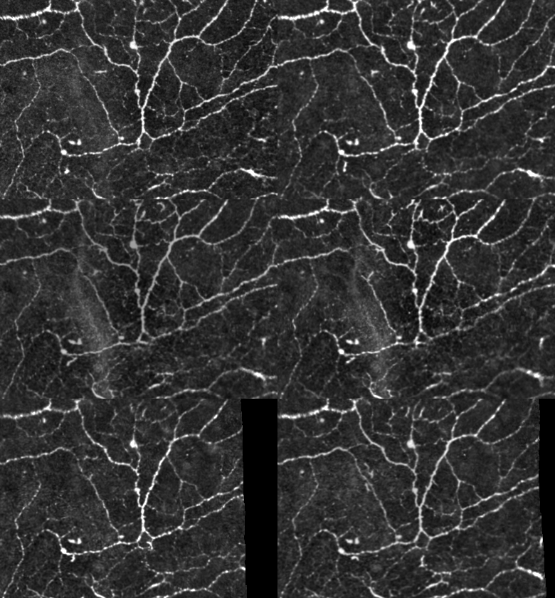
Using our newly developed Fun-IVCM imaging method, we can accurately visualise and identify corneal immune cells in the living human cornea. This novel approach involving dynamic, time-lapsed imaging of corneas, allows us to profile the immune cell identities and behaviours during the early stages of corneal disease. Using confocal microscopy (HRT-RCM, Heidelberg Engineering) at the Lions Eye Institute, cutting-edge imaging of immune cell dynamics is revealing novel insights into human immunology, and accelerating research programs aimed at identifying new markers of corneal and systemic diseases.
In addition, Associate Professor Chinnery and her research program uses preclinical models to investigate fundamental ocular immunology and explore novel therapies for corneal diseases that have an underlying inflammatory basis such as dry eye disease and keratoconus. Ultimately, the aim is to model conditions that affect the human ocular surface to improve and develop new treatments for patients whose sight has been lost to corneal disease, ocular surface diseases or trauma.


